I recently spent a day in (a very rainy) Glasgow with Professor Anne French – RCVS and European Specialist in Veterinary Cardiology. Anne works at Glasgow Vet School where she is Professor of Cardiology and Director of the small animal hospital.
The day was aimed at teaching general practitioners, like myself, to use ultrasound in order to improve our diagnosis and subsequent treatment of heart disease. It was hosted by BCF technologies – one of the foremost manufacturers of veterinary imaging equipment.
As it was a practical course, I had to take my own dog (Oreo) along as my “subject”. He was the model patient and was rewarded with excessive amounts of gravy bones and lots of attention! He was definitely the star of the show- not only for his behaviour – but because all the delegates found his heart easiest to image which is due purely to his size and shape. That was perhaps the first lesson – the size and shape of a dog has a massive influence on the quality of the heart images you are able to get using ultrasound.
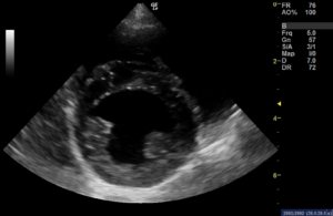
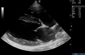
The morning was spent practicing to get the standard cardiac images from which many of our common heart diseases in the dog and cat can be diagnosed. We learned which measurements were important to take and which ultrasound view was necessary to facilitate these measurements. After lunch and a very wet dog walk with the other dogs “helping out” on the course, we repeated the techniques we had learned in the morning. The afternoon session added in the use of colour and spectral Doppler – tools used to investigate the direction and speed of flow of blood within the heart.
It was an excellent and informative course given by a fantastic teacher. Now I need to practice, practice, practice – it’s lucky that Oreo likes gravy bones so much!